1、王小波,王涛.2024年度晚期乳腺癌共识与争议的现状及展望[J].中国癌症杂志,2025,35(3):263-272.
王小波,王涛.2024年度晚期乳腺癌共识与争议的现状及展望[J].中国癌症杂志,2025,35(3):263-272.
2、莫淼,袁晶,周昌明,等.以大型单中心的医院登记为基础的3.5万例乳腺癌患者长期生存报告[J].中国癌症杂志,2020,30(2):90-97.莫淼,袁晶,周昌明,等.以大型单中心的医院登记为基础的3.5万例乳腺癌患者长期生存报告[J].中国癌症杂志,2020,30(2):90-97.
3、吴春晓,庞怡,顾凯,等.2002—2017年上海市女性乳腺癌生存分析[J].中国癌症杂志,2025,35(3):291-297.
吴春晓,庞怡,顾凯,等.2002—2017年上海市女性乳腺癌生存分析[J].中国癌症杂志,2025,35(3):291-297.
4、Huang J J,Chen H X,Li N,et al.Emerging technologies towards extracellular vesicles large-scale production[J].Bioact Mater,2025(52):338-365.Huang J J,Chen H X,Li N,et al.Emerging technologies towards extracellular vesicles large-scale production[J].Bioact Mater,2025(52):338-365.
5、Dixson A C,Dawson T R,Di Vizio D,et al.Context-specific regulation of extracellular vesicle biogenesis and cargo selection[J].Nat Rev Mol Cell Biol,2023,24(7):454-476.
Dixson A C,Dawson T R,Di Vizio D,et al.Context-specific regulation of extracellular vesicle biogenesis and cargo selection[J].Nat Rev Mol Cell Biol,2023,24(7):454-476.
6、Kang Y,Meng L A,Bai S,et al.Extracellular vesicles:The “Trojan Horse” within breast cancer host microenvironments[J].Mol Cancer,2025,24(1):183.Kang Y,Meng L A,Bai S,et al.Extracellular vesicles:The “Trojan Horse” within breast cancer host microenvironments[J].Mol Cancer,2025,24(1):183.
7、王琎,陈建英.细胞外囊泡研究新进展[J].中国组织工程研究,2017,21(4):621-626.王琎,陈建英.细胞外囊泡研究新进展[J].中国组织工程研究,2017,21(4):621-626.
8、Jeppesen D K,Sanchez Z C,Kelley N M,et al.Blebbisomes are large,organelle-rich extracellular vesicles with cell-like properties[J].Nat Cell Biol,2025,27(3):438-448.Jeppesen D K,Sanchez Z C,Kelley N M,et al.Blebbisomes are large,organelle-rich extracellular vesicles with cell-like properties[J].Nat Cell Biol,2025,27(3):438-448.
9、Welsh JA,Goberdhan DCI,O’Driscoll L,et al.Minimal information for studies of extracellular vesicles(MISEV2023):From basic to advanced approaches[J].J Extracell Vesicles,2024,13(2):e12404.Welsh JA,Goberdhan DCI,O’Driscoll L,et al.Minimal information for studies of extracellular vesicles(MISEV2023):From basic to advanced approaches[J].J Extracell Vesicles,2024,13(2):e12404.
10、van Niel G,D’Angelo G,Raposo G.Shedding light on the cell biology of extracellular vesicles[J].Nat Rev Mol Cell Biol,2018,19(4):213-228.van Niel G,D’Angelo G,Raposo G.Shedding light on the cell biology of extracellular vesicles[J].Nat Rev Mol Cell Biol,2018,19(4):213-228.
11、Liu C Q,Liu D X,Wang S,et al.Identification of the SNARE complex that mediates the fusion of multivesicular bodies with the plasma membrane in exosome secretion[J].J Extracellular Vesicle,2023,12(9):12356.Liu C Q,Liu D X,Wang S,et al.Identification of the SNARE complex that mediates the fusion of multivesicular bodies with the plasma membrane in exosome secretion[J].J Extracellular Vesicle,2023,12(9):12356.
12、Perez-Hernandez D,Gutiérrez-Vázquez C,Jorge I,et al.The intracellular interactome of tetraspanin-enriched microdomains reveals their function as sorting machineries toward exosomes[J].J Biol Chem,2013,288(17):11649-11661.
Perez-Hernandez D,Gutiérrez-Vázquez C,Jorge I,et al.The intracellular interactome of tetraspanin-enriched microdomains reveals their function as sorting machineries toward exosomes[J].J Biol Chem,2013,288(17):11649-11661.
13、 Hurwitz S N,Jung S K,Kobulsky D R,et al.Neutral sphingomyelinase blockade enhances hematopoietic stem cell fitness through an integrated stress response[J].Blood,2023,142(20):1708-1723. Hurwitz S N,Jung S K,Kobulsky D R,et al.Neutral sphingomyelinase blockade enhances hematopoietic stem cell fitness through an integrated stress response[J].Blood,2023,142(20):1708-1723.
14、 Hendley A M,AsheE S,Urano A,et al.Abstract A066:NSMase2-mediated exosome secretion shapes the tumor microenvironment to immunologically support pancreatic cancer[J].Cancer Res,2024,84(17_Supplement_2):A066. Hendley A M,AsheE S,Urano A,et al.Abstract A066:NSMase2-mediated exosome secretion shapes the tumor microenvironment to immunologically support pancreatic cancer[J].Cancer Res,2024,84(17_Supplement_2):A066.
15、Liese S,Wenzel E M,Kjos I,et al.Protein crowding mediates membrane remodeling in upstream ESCRT-induced formation of intraluminal vesicles[J].Proc Natl Acad Sci U S A,2020,117(46):28614-28624.
Liese S,Wenzel E M,Kjos I,et al.Protein crowding mediates membrane remodeling in upstream ESCRT-induced formation of intraluminal vesicles[J].Proc Natl Acad Sci U S A,2020,117(46):28614-28624.
16、olf P.The nature and significance of platelet products in human plasma[J].Br J Haematol,1967,13(3):269-288.
olf P.The nature and significance of platelet products in human plasma[J].Br J Haematol,1967,13(3):269-288.
17、Williams J K,Ngo J M,Murugupandiyan A,et al.Calpains orchestrate secretion of annexin-containing microvesicles during membrane repair[J].J Cell Biol,2025,224(7):e202408159.Williams J K,Ngo J M,Murugupandiyan A,et al.Calpains orchestrate secretion of annexin-containing microvesicles during membrane repair[J].J Cell Biol,2025,224(7):e202408159.
18、 Elsharkasy O M,de Voogt W S,Tognoli M L,et al.Integrin beta 1 and fibronectin mediate extracellular vesicle uptake and functional RNA delivery[J].J Biol Chem,2025,301(3):108305.
Elsharkasy O M,de Voogt W S,Tognoli M L,et al.Integrin beta 1 and fibronectin mediate extracellular vesicle uptake and functional RNA delivery[J].J Biol Chem,2025,301(3):108305.
19、Kim Y,Kim J Y,Moon S,et al.Tumor-derived EV miRNA signatures surpass total EV miRNA in supplementing mammography for precision breast cancer diagnosis[J].Theranostics,2024,14(17):6587-6604.Kim Y,Kim J Y,Moon S,et al.Tumor-derived EV miRNA signatures surpass total EV miRNA in supplementing mammography for precision breast cancer diagnosis[J].Theranostics,2024,14(17):6587-6604.
20、Zhai Z,Mu T L,Zhao L N,et al.miR-181a-5p facilitates proliferation,invasion,and glycolysis of breast cancer through NDRG2-mediated activation of PTEN/AKT pathway[J].Bioengineered,2022,13(1):83-95.Zhai Z,Mu T L,Zhao L N,et al.miR-181a-5p facilitates proliferation,invasion,and glycolysis of breast cancer through NDRG2-mediated activation of PTEN/AKT pathway[J].Bioengineered,2022,13(1):83-95.
21、 何建苗,赵华洲,王婷,等.miR-181a-5p靶向Kras对乳腺癌细胞MDA-MB-231增殖及凋亡的调控作用[J].解放军医学杂志,2018,43(9):735-739. 何建苗,赵华洲,王婷,等.miR-181a-5p靶向Kras对乳腺癌细胞MDA-MB-231增殖及凋亡的调控作用[J].解放军医学杂志,2018,43(9):735-739.
22、Li Y Y,Kuscu C,Banach A,et al.miR-181a-5p inhibits cancer cell migration and angiogenesis via downregulation of matrix metalloproteinase-14[J].Cancer Res,2015,75(13):2674-2685.
Li Y Y,Kuscu C,Banach A,et al.miR-181a-5p inhibits cancer cell migration and angiogenesis via downregulation of matrix metalloproteinase-14[J].Cancer Res,2015,75(13):2674-2685.
23、 魏慧君,郭丽丽,李林,等.Warburg效应及其对肿瘤转移的影响[J].中国肺癌杂志,2015,18(3):179-183.
魏慧君,郭丽丽,李林,等.Warburg效应及其对肿瘤转移的影响[J].中国肺癌杂志,2015,18(3):179-183.
24、Colegio O R,Chu N Q,Szabo A L,et al.Functional polarization of tumour-associated macrophages by tumour-derived lactic acid[J].Nature,2014,513(7519):559-563.Colegio O R,Chu N Q,Szabo A L,et al.Functional polarization of tumour-associated macrophages by tumour-derived lactic acid[J].Nature,2014,513(7519):559-563.
25、Liang Y R,Ye F Z,Luo D,et al.Exosomal circSIPA1L3-mediated intercellular communication contributes to glucose metabolic reprogramming and progression of triple negative breast cancer[J].Mol Cancer,2024,23(1):125.Liang Y R,Ye F Z,Luo D,et al.Exosomal circSIPA1L3-mediated intercellular communication contributes to glucose metabolic reprogramming and progression of triple negative breast cancer[J].Mol Cancer,2024,23(1):125.
26、 Sung J S,Kang C W,Kang S,et al.ITGB4-mediated metabolic reprogramming of cancer-associated fibroblasts[J].Oncogene,2020,39(3):664-676. Sung J S,Kang C W,Kang S,et al.ITGB4-mediated metabolic reprogramming of cancer-associated fibroblasts[J].Oncogene,2020,39(3):664-676.
27、Choudhury A,Chatterjee S,Dalui S,et al.Breast cancer cell derived exosomes reduces glycolysis of activated CD8+ T cells in a AKT-mTOR dependent manner[J].Cell Biol Int,2025,49(1):45-54. Choudhury A,Chatterjee S,Dalui S,et al.Breast cancer cell derived exosomes reduces glycolysis of activated CD8+ T cells in a AKT-mTOR dependent manner[J].Cell Biol Int,2025,49(1):45-54.
28、Niu Z X,He J,Wang S Y,et al.Targeting glycolysis for treatment of breast cancer resistance:Current progress and future prospects[J].Int J Biol Sci,2025,21(6):2589-2605.
Niu Z X,He J,Wang S Y,et al.Targeting glycolysis for treatment of breast cancer resistance:Current progress and future prospects[J].Int J Biol Sci,2025,21(6):2589-2605.
29、 Chen J,Cui L K,Lu S T,et al.Amino acid metabolism in tumor biology and therapy[J].Cell Death Dis,2024,15(1):42. Chen J,Cui L K,Lu S T,et al.Amino acid metabolism in tumor biology and therapy[J].Cell Death Dis,2024,15(1):42.
30、Ruan X,Yan W,Cao M,et al.Breast cancer cell-secreted miR-199b-5p hijacks neurometabolic coupling to promote brain metastasis[J].Nat Commun,2024,15(1):4549.
Ruan X,Yan W,Cao M,et al.Breast cancer cell-secreted miR-199b-5p hijacks neurometabolic coupling to promote brain metastasis[J].Nat Commun,2024,15(1):4549.
31、 Li M X,Hu S,Lei H H,et al.Tumor-derived miR-9-5p-loaded EVs regulate cholesterol homeostasis to promote breast cancer liver metastasis in mice[J].Nat Commun,2024,15(1):10539. Li M X,Hu S,Lei H H,et al.Tumor-derived miR-9-5p-loaded EVs regulate cholesterol homeostasis to promote breast cancer liver metastasis in mice[J].Nat Commun,2024,15(1):10539.
32、Ruan X H,Cao M H,Yan W,et al.Cancer‐cell‐secreted extracellular vesicles target p53 to impair mitochondrial function in muscle[J].EMBO Rep,2023,24(9):EMBR202256464.Ruan X H,Cao M H,Yan W,et al.Cancer‐cell‐secreted extracellular vesicles target p53 to impair mitochondrial function in muscle[J].EMBO Rep,2023,24(9):EMBR202256464.
33、Xie Y F,Liu F F,Wu Y F,et al.Inflammation in cancer:Therapeutic opportunities from new insights[J].Mol Cancer,2025,24(1):51.Xie Y F,Liu F F,Wu Y F,et al.Inflammation in cancer:Therapeutic opportunities from new insights[J].Mol Cancer,2025,24(1):51.
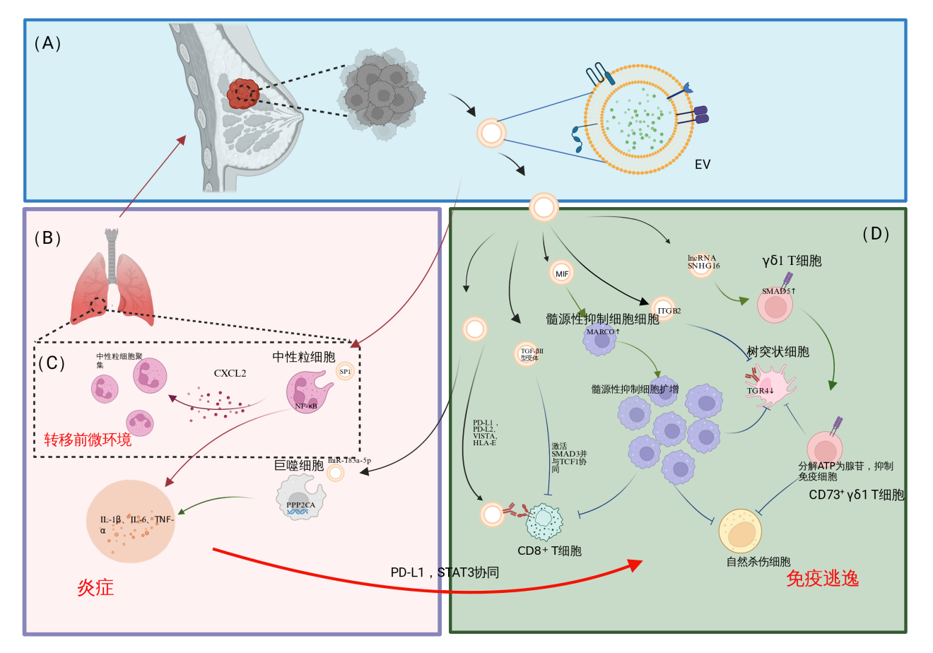
34、Liu S J,Tian B L,Wang N,et al.MARCO expression on myeloid-derived suppressor cells is essential for their differentiation and immunosuppression[J].Cell Death Discov,2025,11(1):337.Liu S J,Tian B L,Wang N,et al.MARCO expression on myeloid-derived suppressor cells is essential for their differentiation and immunosuppression[J].Cell Death Discov,2025,11(1):337.
35、Paierhati P,Ma B,Abudukeremu M.Exosomal ITGB2 mediates immune evasion in triple-negative breast cancer by suppressing dendritic cell activation via TLR4[J].Iranian Journal of Public Health,2025,54(6):1252-1262.Paierhati P,Ma B,Abudukeremu M.Exosomal ITGB2 mediates immune evasion in triple-negative breast cancer by suppressing dendritic cell activation via TLR4[J].Iranian Journal of Public Health,2025,54(6):1252-1262.
36、 Ni C,Fang Q Q,Chen W Z,et al.Breast cancer-derived exosomes transmit lncRNA SNHG16 to induce CD73+γδ1 Treg cells[J].Sig Transduct Target Ther,2020,5(1):41.
Ni C,Fang Q Q,Chen W Z,et al.Breast cancer-derived exosomes transmit lncRNA SNHG16 to induce CD73+γδ1 Treg cells[J].Sig Transduct Target Ther,2020,5(1):41.
37、杜晓月,温少迪,沈波.肿瘤来源的外泌体PD-L1在肿瘤中的研究进展[J].中国肿瘤外科杂志,2022,14(5):515-520.杜晓月,温少迪,沈波.肿瘤来源的外泌体PD-L1在肿瘤中的研究进展[J].中国肿瘤外科杂志,2022,14(5):515-520.
38、Xie F,Zhou X X,Su P,et al.Breast cancer cell-derived extracellular vesicles promote CD8+ T cell exhaustion via TGF-β type II receptor signaling[J].Nat Commun,2022,13(1):4461.Xie F,Zhou X X,Su P,et al.Breast cancer cell-derived extracellular vesicles promote CD8+ T cell exhaustion via TGF-β type II receptor signaling[J].Nat Commun,2022,13(1):4461.
39、Qiu Y F,Yang Y,Yang R Y,et al.Activated T cell-derived exosomal PD-1 attenuates PD-L1-induced immune dysfunction in triple-negative breast cancer[J].Oncogene,2021,40(31):4992-5001.
Qiu Y F,Yang Y,Yang R Y,et al.Activated T cell-derived exosomal PD-1 attenuates PD-L1-induced immune dysfunction in triple-negative breast cancer[J].Oncogene,2021,40(31):4992-5001.
40、 Zhang L Y,Pan J,Wang M J,et al.Chronic stress-induced and tumor derived SP1+ exosomes polarizing IL-1β+ neutrophils to increase lung metastasis of breast cancer[J].Adv Sci,2025,12(4):2310266. Zhang L Y,Pan J,Wang M J,et al.Chronic stress-induced and tumor derived SP1+ exosomes polarizing IL-1β+ neutrophils to increase lung metastasis of breast cancer[J].Adv Sci,2025,12(4):2310266.
41、Guo J,Duan Z J,Zhang C,et al.Mouse 4T1 breast cancer cell–derived exosomes induce proinflammatory cytokine production in macrophages via miR-183[J].J Immunol,2020,205(10):2916-2925.
Guo J,Duan Z J,Zhang C,et al.Mouse 4T1 breast cancer cell–derived exosomes induce proinflammatory cytokine production in macrophages via miR-183[J].J Immunol,2020,205(10):2916-2925.
42、Thuya W L,Cao Y,Ho P C,et al.Insights into IL-6/JAK/STAT3 signaling in the tumor microenvironment:Implications for cancer therapy[J].Cytokine Growth Factor Rev,2025(85):26-42.
Thuya W L,Cao Y,Ho P C,et al.Insights into IL-6/JAK/STAT3 signaling in the tumor microenvironment:Implications for cancer therapy[J].Cytokine Growth Factor Rev,2025(85):26-42.
43、 Sun X T,Wang X R,Yan C G,et al.Tumor cell-released LC3-positive EVs promote lung metastasis of breast cancer through enhancing premetastatic niche formation[J].Cancer Sci,2022,113(10):3405-3416. Sun X T,Wang X R,Yan C G,et al.Tumor cell-released LC3-positive EVs promote lung metastasis of breast cancer through enhancing premetastatic niche formation[J].Cancer Sci,2022,113(10):3405-3416.
44、Hoshino A,Costa-Silva B,Shen T L,et al.Tumour exosome integrins determine organotropic metastasis[J].Nature,2015,527(7578):329-335.Hoshino A,Costa-Silva B,Shen T L,et al.Tumour exosome integrins determine organotropic metastasis[J].Nature,2015,527(7578):329-335.
45、 Lv Y,Li Y,Zhou J,et al.Exosomal miR-122-5p for regulation of secretory functions of fibroblasts and promotion of breast cancer metastasis by targeting MKP-2:An experimental study[J].Cancer Biol Ther,2025,26(1):2500104. Lv Y,Li Y,Zhou J,et al.Exosomal miR-122-5p for regulation of secretory functions of fibroblasts and promotion of breast cancer metastasis by targeting MKP-2:An experimental study[J].Cancer Biol Ther,2025,26(1):2500104.
46、 Arnold J,Schattschneider J,Blechner C,et al.Tubulin Tyrosine Ligase Like 4(TTLL4) overexpression in breast cancer cells is associated with brain metastasis and alters exosome biogenesis[J].J Exp Clin Cancer Res,2020,39(1):205. Arnold J,Schattschneider J,Blechner C,et al.Tubulin Tyrosine Ligase Like 4(TTLL4) overexpression in breast cancer cells is associated with brain metastasis and alters exosome biogenesis[J].J Exp Clin Cancer Res,2020,39(1):205.
47、Liu X Y,Ma R,Wei F,et al.Tumor-derived exosomal lncRNA-MIR193BHG promotes bone metastasis of breast cancer by targeting the miR-489-3p/DNMT3A signaling axis in osteoclasts[J].J Transl Med,2025,23(1):142.
Liu X Y,Ma R,Wei F,et al.Tumor-derived exosomal lncRNA-MIR193BHG promotes bone metastasis of breast cancer by targeting the miR-489-3p/DNMT3A signaling axis in osteoclasts[J].J Transl Med,2025,23(1):142.
48、Hao C,Sheng Z M,Wang W H,et al.Tumor-derived exosomal miR-148b-3p mediates M2 macrophage polarization via TSC2/mTORC1 to promote breast cancer migration and invasion[J].Thorac Cancer,2023,14(16):1477-1491.
Hao C,Sheng Z M,Wang W H,et al.Tumor-derived exosomal miR-148b-3p mediates M2 macrophage polarization via TSC2/mTORC1 to promote breast cancer migration and invasion[J].Thorac Cancer,2023,14(16):1477-1491.
49、 Li H T,Fan J J,Ma J,et al.Exosomal miR-182-5p from breast cancer cells reprogram tumor-associated macrophages and promote triple-negative breast cancer progression by targeting Notch1 in macrophages[J].Archives of Medical Science,2025,21(4):1471-1486.
Li H T,Fan J J,Ma J,et al.Exosomal miR-182-5p from breast cancer cells reprogram tumor-associated macrophages and promote triple-negative breast cancer progression by targeting Notch1 in macrophages[J].Archives of Medical Science,2025,21(4):1471-1486.
50、Chai Y M,Shi Y.Exosomal POU5 F1 derived from TNBC promotes cancer progression by regulating M2 macrophage polarization via inhibiting TRAF6 ubiquitination and activating AKT in macrophage[J].Cell Biol Toxicol,2025,41(1):95.Chai Y M,Shi Y.Exosomal POU5 F1 derived from TNBC promotes cancer progression by regulating M2 macrophage polarization via inhibiting TRAF6 ubiquitination and activating AKT in macrophage[J].Cell Biol Toxicol,2025,41(1):95.
51、Wang Z Y,Zhang C,Guo J,et al.Exosomal miRNA-223-3p derived from tumor associated macrophages promotes pulmonary metastasis of breast cancer 4T1 cells[J].Transl Oncol,2023(35):101715.
Wang Z Y,Zhang C,Guo J,et al.Exosomal miRNA-223-3p derived from tumor associated macrophages promotes pulmonary metastasis of breast cancer 4T1 cells[J].Transl Oncol,2023(35):101715.
52、Chen F,Chen J N,Yang L B,et al.Extracellular vesicle-packaged HIF-1α-stabilizing lncRNA from tumour-associated macrophages regulates aerobic glycolysis of breast cancer cells[J].Nat Cell Biol,2019,21(4):498-510.
Chen F,Chen J N,Yang L B,et al.Extracellular vesicle-packaged HIF-1α-stabilizing lncRNA from tumour-associated macrophages regulates aerobic glycolysis of breast cancer cells[J].Nat Cell Biol,2019,21(4):498-510.
53、 Huang S L,Fan P Z,Zhang C J,et al.Exosomal microRNA-503-3p derived from macrophages represses glycolysis and promotes mitochondrial oxidative phosphorylation in breast cancer cells by elevating DACT2[J].Cell Death Discov,2021, 7(1):119.
Huang S L,Fan P Z,Zhang C J,et al.Exosomal microRNA-503-3p derived from macrophages represses glycolysis and promotes mitochondrial oxidative phosphorylation in breast cancer cells by elevating DACT2[J].Cell Death Discov,2021, 7(1):119.
54、Yue S J,Ye X S,Zhou T,et al.PGRN-/- TAMs-derived exosomes inhibit breast cancer cell invasion and migration and its mechanism exploration[J].Life Sci,2021(264):118687.
Yue S J,Ye X S,Zhou T,et al.PGRN-/- TAMs-derived exosomes inhibit breast cancer cell invasion and migration and its mechanism exploration[J].Life Sci,2021(264):118687.
55、Castagnoli L,Franceschini A,Cancila V,et al.CD36 enrichment in HER2-positive mesenchymal stem cells drives therapy refractoriness in breast cancer[J].J Exp Clin Cancer Res,2025,44(1):19.Castagnoli L,Franceschini A,Cancila V,et al.CD36 enrichment in HER2-positive mesenchymal stem cells drives therapy refractoriness in breast cancer[J].J Exp Clin Cancer Res,2025,44(1):19.
56、Zhu Q,Cao Y R,Yuan J Q,et al.Adipose-derived stem cell exosomes promote tumor characterization and immunosuppressive microenvironment in breast cancer[J].Cancer Immunol Immunother,2024,73(2):39.
Zhu Q,Cao Y R,Yuan J Q,et al.Adipose-derived stem cell exosomes promote tumor characterization and immunosuppressive microenvironment in breast cancer[J].Cancer Immunol Immunother,2024,73(2):39.
57、 Li T,Zhou X H,Wang J Z,et al.Adipose-derived mesenchymal stem cells and extracellular vesicles confer antitumor activity in preclinical treatment of breast cancer[J].Pharmacol Res,2020(157):104843.
Li T,Zhou X H,Wang J Z,et al.Adipose-derived mesenchymal stem cells and extracellular vesicles confer antitumor activity in preclinical treatment of breast cancer[J].Pharmacol Res,2020(157):104843.
58、Du L,Tao X G,Shen X W.Human umbilical cord mesenchymal stem cell-derived exosomes inhibit migration and invasion of breast cancer cells via miR-21-5p/ZNF367 pathway[J].Breast Cancer,2021,28(4):829-837.
Du L,Tao X G,Shen X W.Human umbilical cord mesenchymal stem cell-derived exosomes inhibit migration and invasion of breast cancer cells via miR-21-5p/ZNF367 pathway[J].Breast Cancer,2021,28(4):829-837.
59、Luo T,Liu Q Y,Tan A H,et al.Mesenchymal stem cell-secreted exosome promotes chemoresistance in breast cancer via enhancing miR-21-5p-mediated S100A6 expression[J].Mol Ther Oncolytics,2020(19):283-293.
Luo T,Liu Q Y,Tan A H,et al.Mesenchymal stem cell-secreted exosome promotes chemoresistance in breast cancer via enhancing miR-21-5p-mediated S100A6 expression[J].Mol Ther Oncolytics,2020(19):283-293.
60、 Zhang Y,Lai X F,Yue Q F,et al.Bone marrow mesenchymal stem cells-derived exosomal microRNA-16-5p restrains epithelial-mesenchymal transition in breast cancer cells via EPHA1/NF-κB signaling axis[J].Genomics,2022,114(3):110341. Zhang Y,Lai X F,Yue Q F,et al.Bone marrow mesenchymal stem cells-derived exosomal microRNA-16-5p restrains epithelial-mesenchymal transition in breast cancer cells via EPHA1/NF-κB signaling axis[J].Genomics,2022,114(3):110341.
61、Jafari N,Kolla M,Meshulam T,et al.Adipocyte-derived exosomes may promote breast cancer progression in type 2 diabetes[J].Sci Signal,2021,14(710):eabj2807.Jafari N,Kolla M,Meshulam T,et al.Adipocyte-derived exosomes may promote breast cancer progression in type 2 diabetes[J].Sci Signal,2021,14(710):eabj2807.
62、 Li C,Qiu S,Liu X H,et al.Extracellular matrix-derived mechanical force governs breast cancer cell stemness and quiescence transition through integrin-DDR signaling[J].Signal Transduct Target Ther,2023,8(1):247. Li C,Qiu S,Liu X H,et al.Extracellular matrix-derived mechanical force governs breast cancer cell stemness and quiescence transition through integrin-DDR signaling[J].Signal Transduct Target Ther,2023,8(1):247.
63、Zhao D,Wu K,Sharma S,et al.Exosomal miR-1304-3p promotes breast cancer progression in African Americans by activating cancer-associated adipocytes[J].Nat Commun,2022(13):7734.
Zhao D,Wu K,Sharma S,et al.Exosomal miR-1304-3p promotes breast cancer progression in African Americans by activating cancer-associated adipocytes[J].Nat Commun,2022(13):7734.
64、Wu X Y,Wang S Y,Lin L,et al.Association between triglyceride glucose index and breast cancer in 142,184 Chinese adults:Findings from the REACTION study[J].Front Endocrinol,2024(15):1321622.
Wu X Y,Wang S Y,Lin L,et al.Association between triglyceride glucose index and breast cancer in 142,184 Chinese adults:Findings from the REACTION study[J].Front Endocrinol,2024(15):1321622.
65、周文重.肥胖通过脂肪细胞分泌因子和外泌体促进乳腺癌转移的分子机制研究[D].济南:山东大学,2020.周文重.肥胖通过脂肪细胞分泌因子和外泌体促进乳腺癌转移的分子机制研究[D].济南:山东大学,2020.
66、 Li Y H,Jiang B H,Zeng L J,et al.Adipocyte-derived exosomes promote the progression of triple-negative breast cancer through circCRIM1-dependent OGA activation[J].Environ Res,2023,239(Pt 1):117266.
Li Y H,Jiang B H,Zeng L J,et al.Adipocyte-derived exosomes promote the progression of triple-negative breast cancer through circCRIM1-dependent OGA activation[J].Environ Res,2023,239(Pt 1):117266.
67、王文浩.乳腺癌细胞来源外泌体miR-21-5p靶向NF2诱导成纤维细胞转化为CAFs的机制研究[D].济南:山东大学,2024.
王文浩.乳腺癌细胞来源外泌体miR-21-5p靶向NF2诱导成纤维细胞转化为CAFs的机制研究[D].济南:山东大学,2024.
68、de Luca G,Petrillo G,Scognamiglio I,et al.Fibroblasts activated by miRs-185-5p,miR-652-5p,and miR-1246 shape the tumor microenvironment in triple-negative breast cancer via PATZ1 downregulation[J].Cell Mol Life Sci,2025,82(1):287.de Luca G,Petrillo G,Scognamiglio I,et al.Fibroblasts activated by miRs-185-5p,miR-652-5p,and miR-1246 shape the tumor microenvironment in triple-negative breast cancer via PATZ1 downregulation[J].Cell Mol Life Sci,2025,82(1):287.
69、Ye F Z,Liang Y R,Wang Y J,et al.Cancer-associated fibroblasts facilitate breast cancer progression through exosomal circTBPL1-mediated intercellular communication[J].Cell Death Dis,2023(14):471.Ye F Z,Liang Y R,Wang Y J,et al.Cancer-associated fibroblasts facilitate breast cancer progression through exosomal circTBPL1-mediated intercellular communication[J].Cell Death Dis,2023(14):471.
70、Chen B,Sang Y T,Song X J,et al.Exosomal miR-500a-5p derived from cancer-associated fibroblasts promotes breast cancer cell proliferation and metastasis through targeting USP28[J].Theranostics,2021,11(8):3932-3947.Chen B,Sang Y T,Song X J,et al.Exosomal miR-500a-5p derived from cancer-associated fibroblasts promotes breast cancer cell proliferation and metastasis through targeting USP28[J].Theranostics,2021,11(8):3932-3947.
71、Tao S,Gao Y L,Wang X,et al.CAF-derived exosomal LINC01711 promotes breast cancer progression by activating the miR-4510/NELFE axis and enhancing glycolysis[J].FASEB J,2025,39(7):e70471.
Tao S,Gao Y L,Wang X,et al.CAF-derived exosomal LINC01711 promotes breast cancer progression by activating the miR-4510/NELFE axis and enhancing glycolysis[J].FASEB J,2025,39(7):e70471.
72、 Sheng Z M,Wang X J,Ding X D,et al.Exosomal miRNA-92a derived from cancer-associated fibroblasts promote invasion and metastasis in breast cancer by regulating G3BP2[J].Cell Signal,2024(119):111182.
Sheng Z M,Wang X J,Ding X D,et al.Exosomal miRNA-92a derived from cancer-associated fibroblasts promote invasion and metastasis in breast cancer by regulating G3BP2[J].Cell Signal,2024(119):111182.
73、Li X Y,M?ller S H,Park J,et al.Tumor-instructed glutamine synthesis in cancer-associated fibroblasts promotes pro-tumor macrophages[J].J Exp Med,2025,222(9):e20241426.
Li X Y,M?ller S H,Park J,et al.Tumor-instructed glutamine synthesis in cancer-associated fibroblasts promotes pro-tumor macrophages[J].J Exp Med,2025,222(9):e20241426.
74、Cheytan T,Schneider M,Würth R,et al.Small extracellular vesicles and particles(sEVPs) derived from tumor-free pre-metastatic organs promote breast cancer metastasis and support organotropism[J].Mol Cancer,2025,24(1):72.
Cheytan T,Schneider M,Würth R,et al.Small extracellular vesicles and particles(sEVPs) derived from tumor-free pre-metastatic organs promote breast cancer metastasis and support organotropism[J].Mol Cancer,2025,24(1):72.
75、Gounis M,Campos AV,Shokry E,et al.Metabolic adaptations of micrometastases alter EV production to generate invasive microenvironments[J].J Cell Biol,2025,224(8):e202405061.Gounis M,Campos AV,Shokry E,et al.Metabolic adaptations of micrometastases alter EV production to generate invasive microenvironments[J].J Cell Biol,2025,224(8):e202405061.
76、 Omarini C,Catani V,Mastrolia I,et al.Extracellular vesicles-derived miR-21 as a biomarker for early diagnosis and tumor activity in breast cancer subtypes[J].Biomark Res,2025,13(1):14.
Omarini C,Catani V,Mastrolia I,et al.Extracellular vesicles-derived miR-21 as a biomarker for early diagnosis and tumor activity in breast cancer subtypes[J].Biomark Res,2025,13(1):14.
77、Moon P G,Lee J E,Cho Y E,et al.Identification of developmental endothelial locus-1 on circulating extracellular vesicles as a novel biomarker for early breast cancer detection[J].Clin Cancer Res,2016,22(7):1757-1766.Moon P G,Lee J E,Cho Y E,et al.Identification of developmental endothelial locus-1 on circulating extracellular vesicles as a novel biomarker for early breast cancer detection[J].Clin Cancer Res,2016,22(7):1757-1766.
78、Park M,Lee C H,Noh H,et al.High-precision extracellular-vesicle isolation-analysis integrated platform for rapid cancer diagnosis directly from blood plasma[J].Biosens Bioelectron,2025,267:116863.Park M,Lee C H,Noh H,et al.High-precision extracellular-vesicle isolation-analysis integrated platform for rapid cancer diagnosis directly from blood plasma[J].Biosens Bioelectron,2025,267:116863.
79、Shie M Y,Huang S W,Chen Y,et al.Engineering HLA-G-targeted extracellular vesicles nanoplatform for enhanced cancer therapy through precise cancer drug delivery[J].Nat Commun,2025,16(1):11308.
Shie M Y,Huang S W,Chen Y,et al.Engineering HLA-G-targeted extracellular vesicles nanoplatform for enhanced cancer therapy through precise cancer drug delivery[J].Nat Commun,2025,16(1):11308.
80、Kalluri V S,Smaglo B G,Mahadevan K K,et al.Engineered exosomes with KrasG12D specific siRNA in pancreatic cancer:A phase I study with immunological correlates[J].Nat Commun,2025,16(1):8696.
Kalluri V S,Smaglo B G,Mahadevan K K,et al.Engineered exosomes with KrasG12D specific siRNA in pancreatic cancer:A phase I study with immunological correlates[J].Nat Commun,2025,16(1):8696.